Models must identify what changed, where it changed, and how that change evolves across timepoints.
Real-world radiology is comparative and longitudinal: disease progression is assessed by comparing current and prior MRI scans across multiple anatomical views and sequences. The Time-Aware Multi-View MRI Benchmark evaluates whether foundation models can reason over these longitudinal changes rather than interpret isolated images. It brings together 3,920 expert-verified question-answer pairs from 890 patients and more than 3,200 MRI timepoints across seven longitudinal cohorts, spanning glioblastoma, brain metastases, neurodegeneration, and vestibular schwannoma. Across 16 vision-language models, the benchmark reveals persistent limitations in clinically important change characterization, temporal reasoning, and quantitative assessment.
Models must identify what changed, where it changed, and how that change evolves across timepoints.
Axial, coronal, and sagittal evidence is evaluated together rather than treating scans as isolated 2D images.
The benchmark spans multiple diseases and uses expert-reviewed question-answer pairs.

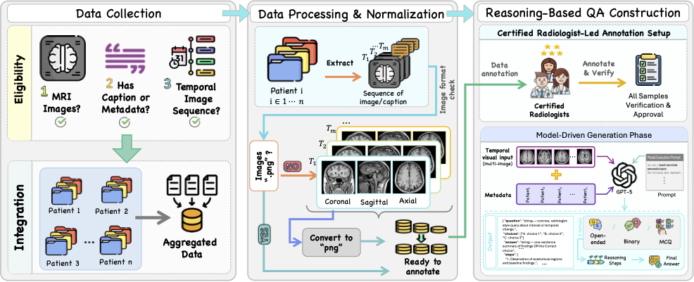
Benchmark construction pipeline. Longitudinal MRI cohorts are harmonized through multi-view extraction and preprocessing, followed by expert-guided QA generation and radiologist verification.
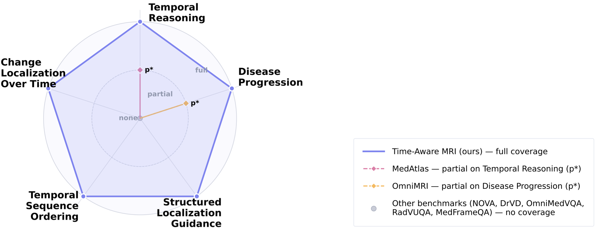
The benchmark evaluates foundation models on five complementary tasks designed around longitudinal MRI interpretation and disease progression reasoning.
Temporal Reasoning · Disease Progression · Structured Localization Guidance · Temporal Sequence Ordering · Change Localization Over Time

Comparison with existing MRI benchmarks. Time-Aware MRI provides full coverage across all five reasoning dimensions.
Interpret interval changes across sequential scans, planes, and contrasts.
Characterize growth, regression, or stability over time.
Identify changed anatomy, boundaries, features, and confounders.
Arrange scans chronologically from visible progression patterns.
Identify the timepoint and spatial region showing maximal change.

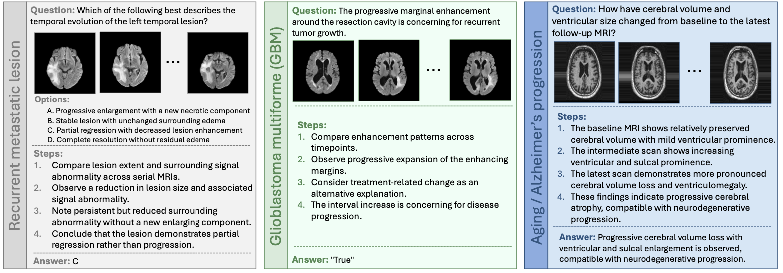
Representative longitudinal MRI reasoning samples across metastatic lesions, glioblastoma, and neurodegenerative progression.
Across 16 vision-language models, current systems achieve moderate temporal alignment but remain limited in clinically important change characterization and quantitative reasoning.
InternVL3.5-Inst
InternVL3.5-Inst
Peak result in the multi-view analysis
Across evaluated models
Source MRI images are not redistributed through this project page or repository. Researchers should obtain the source datasets from their official providers and follow the corresponding licenses and data-use agreements. The repository provides preprocessing and evaluation code for reproducing the benchmark workflow.
@inproceedings{alghallabi2026timeaware,
title={How Good are Foundation Models in Longitudinal MRI Disease Progression Reasoning?},
author={Al Ghallabi, Wafa and Thawkar, Ritesh and Ghaboura, Sara and Thawakar, Omkar and Saeed, Numan and Al Nuaimi, Dana and Alkatheeri, Ajnas and Khan, Salman and Khan, Fahad Shahbaz},
booktitle={Medical Image Computing and Computer Assisted Intervention -- MICCAI 2026},
year={2026}
}